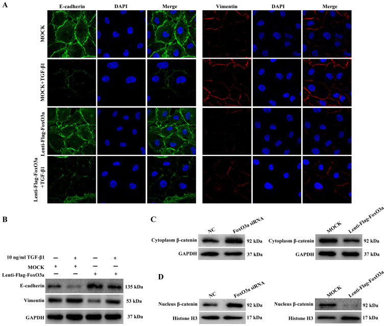
Figure 5.
FoxO3a suppresses TGF-β1-induced EMT and β-catenin expression. (A) Immunofluorescence (magnification, ×400) and (B) western blot analysis were performed to detect the expression of TGF-β1-induced EMT markers with FoxO3a overexpression. (C) Western blot analysis was performed to detect the cytoplasmic expression of β-catenin with either FoxO3a knockdown (left panel) or overexpression (right panel). (D) Western blot analysis was performed to verify the changes in β-catenin levels in the nucleus with either FoxO3a knockdown (left panel) or overexpression (right panel). The nuclei protein was normalized to Histone H3. EMT, epithelial-mesenchymal transition; FoxO3a, Forkhead box O3a; TGF-β1, transforming growth factor-β1; siRNA, small interfering RNA; NC, negative control.

