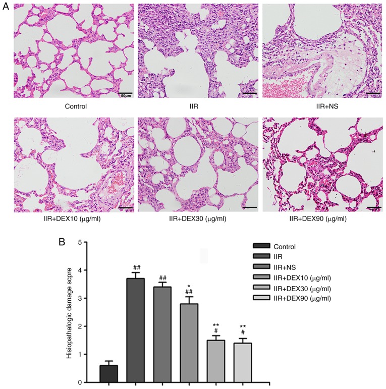
Figure 1.
Histopathological changes in the lung tissue of control, IIR, IIR + NS, IIR + DEX (10 µg/kg), IIR + DEX (30 µg/kg) and IIR + DEX (90 µg/kg) groups. (A) Tissue sections of the lung tissue stained with hematoxylin and eosin in different groups. Magnification, ×40. (B) Pathological damage scores of the lung tissue in the control, IIR + NS, IIR + DEX (10 µg/kg), IIR + DEX (30 µg/kg) and IIR + DEX (90 µg/kg) groups. Data are presented as the mean ± standard error of the mean. #P<0.05 vs. control group; ##P<0.01 vs. control group; *P<0.05 vs. IIR + NS group; **P<0.01 vs. IIR + NS group. IIR, intestinal ischemia reperfusion; DEX, dexmedetomidine; NS, normal saline.

