Figure 1.

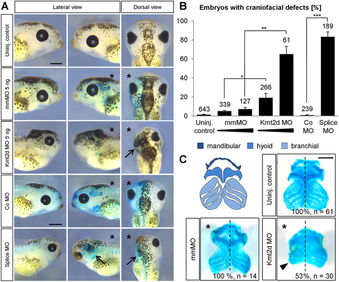
Kmt2d loss-of-function causes craniofacial malformations. (A) Phenotypic analysis of craniofacial defects at stage 42. Embryos were injected with 2.5–5 ng of mmMO or Kmt2d MO or 5 ng control (Co) MO or splice MO into one blastomere at the two-cell stage. 150 pg lacZ mRNA was co-injected as a lineage tracer. The injected side of the embryo is marked (asterisk). Both the Kmt2d MO and the splice MO cause severe craniofacial malformations (arrows), compared to mmMO- and Co MO-treated controls. Scale bars = 500 μm. (B) Graph summarizing the results from at least four independent experiments; ±SEM and the number of analyzed embryos are given for each condition. A two-tailed unpaired Student’s t-test was applied. (C) Cartilage staining. Schematic representation of the cranial cartilage structures of a Xenopus laevis embryo (upper left panel). Embryos were injected with 2.5 ng mmMO or Kmt2d MO in combination with 50 pg mGFP mRNA as a lineage tracer into one blastomere at the two-cell stage, and the cranial cartilage was visualized by Alcian Blue staining at stage 44. Kmt2d knockdown leads to reduced cranial cartilage (arrowhead) on the injected side (asterisk). Scale bar = 500 μm.
