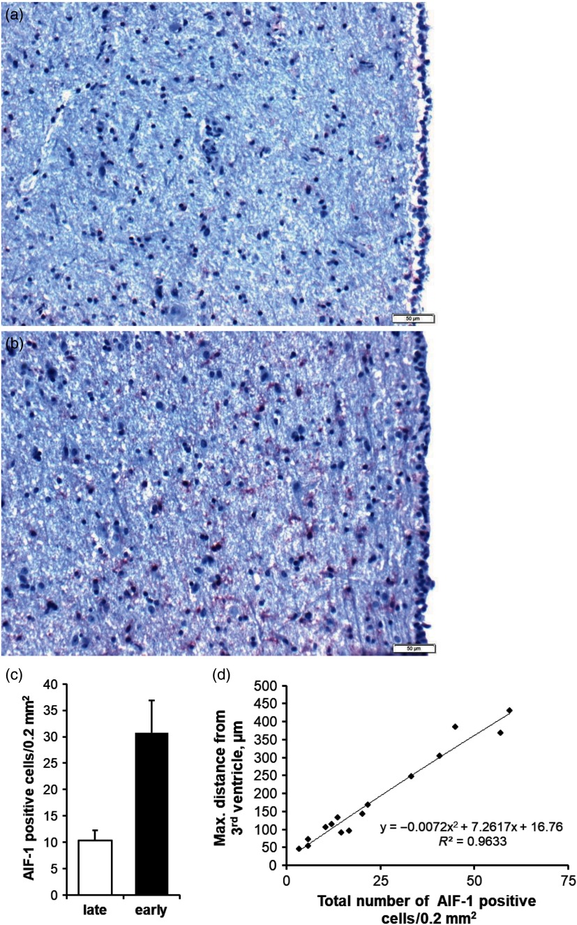
Figure 2.
Microglia activation in the periventricular region of the hypothalamus of late- (a) and early-lactating (b) dairy cows. Activated microglia was immunostained for allograft inflammatory factor 1 (AIF-1) and visualised in red; cell nuclei were counterstained by haematoxylin (blue). Epithelial cells of the third ventricle are located at the right image margins. The scale bar indicates 50 µm. The number of activated microglia in the periventricular hypothalamic region in up to 500 µm radial distance from the third ventricle epithelium during early and late lactation is given as mean ± SE (c). Exponential relationship and coefficient of determination (R2) between the maximal distance of an AIF-1-positive cell from the third ventricle border and the total number of AIF-1-positive cells (d). Adapted from B. Kuhla (unpublished data).

