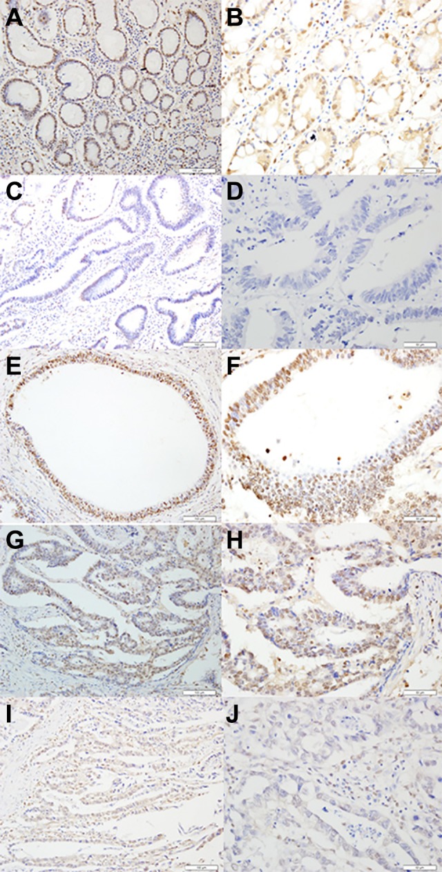
Figure 1.

Representative of SRY-related HMG box-12 (SOX12) expression in adjacent nontumor tissues and primary gastric cancer tissues detected by immunostaining with anti-SOX12 antibody (×200 or ×400) .The evaluation was based on the staining intensity and extent of staining. Staining intensity was scored as 0 (negative), 1 (weak), 2 (moderate), and 3 (strong). A and B, Staining status of adjacent nontumor tissues (strong staining). C and D, Staining of SOX12 in gastric cancer tissues (negative). E and F, Staining of SOX12 in gastric cancer tissues (strong staining). G and H, Staining of SOX12 in gastric cancer tissues (medium staining). I and J, Staining of SOX12 in gastric cancer tissues (weak staining).
