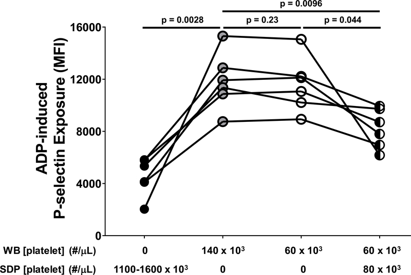
Figure 5. Single donor platelet (SDP) units reduced the responsiveness of platelets in fresh whole blood to activation by adenosine diphosphate (ADP).
Apheresis-derived SDP units were obtained from Versiti – Blood Center of Wisconsin and left unmodified (black circles). Whole blood (WB) samples were obtained from healthy adult volunteers and manipulated to achieve a normal platelet count (140 × 103/μL; gray circles) or were made thrombocytopenic to approximate platelet counts observed in neonates on cardiopulmonary bypass (60 × 103/μL; open circles). SDP units were mixed with thrombocytopenic WB samples to achieve a final platelet count of 140 × 103/μL, with 60 × 103/μL platelets derived from WB and 80 × 103/μL platelets derived from SDP units (black and white circles). Each of these platelet preparations was stimulated with ADP (20 μM). Activated platelets were identified by flow cytometry following staining with a phycoerythrin (PE)-tagged antibody specific for the platelet α-granule constituent, P-selectin. Results are reported as PE median fluorescence intensity (MFI). Each symbol represents the result for a single experiment and lines represent relationships between SDP and WB samples studied in a given experiment (n=6). Because the data were normally distributed, the paired t-test was used to compare thrombocytopenic whole blood samples to those with normal platelet counts. Statistically significant differences between groups are indicated by p values. Note that, compared to WB with a platelet count of 140 × 103/μL, the response to stimulation with ADP was lower in SDP units, the same in WB samples with a thrombocytopenic platelet count of 60 × 103/μL, and reduced in thrombocytopenic WB samples to which platelets from SDP units were added to achieve a platelet count of 140 × 103/μL.

