
Figure 18.
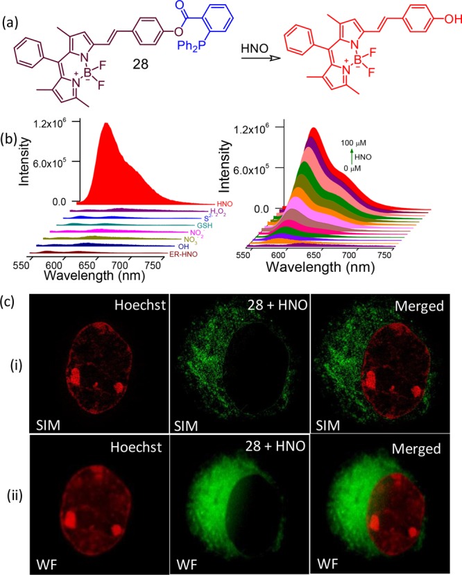
(a) Molecular structure of the reagent 28, (b) luminescence spectral profile to show the specificity of the reagent toward 28 and its concentration-dependent intensity variation, (c) dual-color (i) SIM and (ii) comparative wide-field CLSM images with Hoechst as the nuclear stain (pseudo coloring has been employed in all the images) and 28 as the ER-specific stain. Figure 18(b,c) was reprinted from ref (47). Copyright 2017. American Chemical Society.