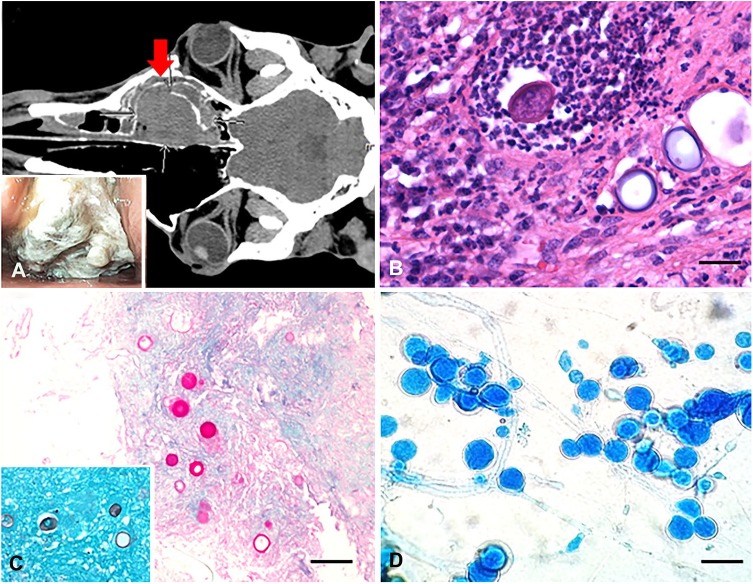
Figure 1.
An obstructive nasal pyogranuloma caused by Flavodon flavus infection in a horse. A. Dorsal plane computed tomography image of the head (soft tissue algorithm) demonstrates the large, soft tissue–attenuating, partially mineralized mass (arrow) occupying the width of the right nasal passage and dorsal-conchofrontal sinus. Inset: nasal passage endoscopy shows a large obstructive irregular mass covered with necropurulent exudate. B. Multiple spherical fungal structures admixed with many degenerate neutrophils and other inflammatory cells. H&E. Bar = 80 µm. C. The wall of the round fungal structures is periodic acid–Schiff stain positive and Grocott methenamine silver stain positive (inset). Bar = 200 µm. D. Lactophenol cotton blue stain of the fungal colonies identified numerous conidia in clusters, associated with parallel-walled and branching hyphae. Bar = 150 µm.

