
Fig. 1.
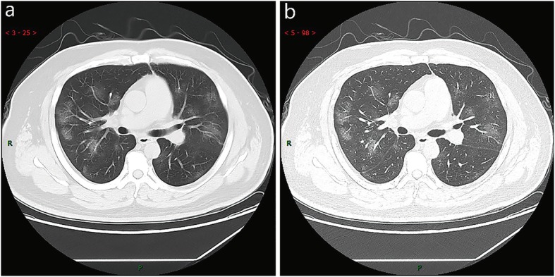
Typical CT imaging manifestation (case 1). A 38 years old male with fever without obvious inducement (39.3 ℃), dry cough and shortness of breath for 3 days. Laboratory test: normal white blood cells (6.35 × 10⁹/L), decreased lymphocytes percentage (4.1%), decreased lymphocytes count (0.31 × 10⁹/L), decreased eosinophil count (0 × 109/L)), increased C-reaction protein (170.91 mg/L), increased procalcitonin (0.45 ng/ml). Imaging examination: multiple patches, grid-like lobule and thickening of interlobular septa, typical “paving stone-like” signs. a SL(Slice): 6 mm; b high-resolution computed tomography(HRCT). HRCT. high-resolution computed tomography