
Fig. 7.
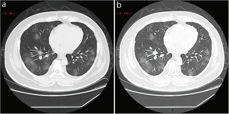
CT imaging of early stage. Male, 38 years old, fever without obvious inducement (39.3 ℃), dry cough and shortness of breath for 3 days. Laboratory test: decreased white blood cells (3.01 × 10⁹/L), decreased lymphocytes (0.81 × 10⁹/ L), increased C-reaction protein (60.8 mg/L), increased procalcitonin (0.16 ng/ml). Imaging examination: a (thin layer CT) and b (high-resolution CT) showed multiple patchy and light consolidation in both lungs and grid-like thickness of interlobular septa