
Figure A1.
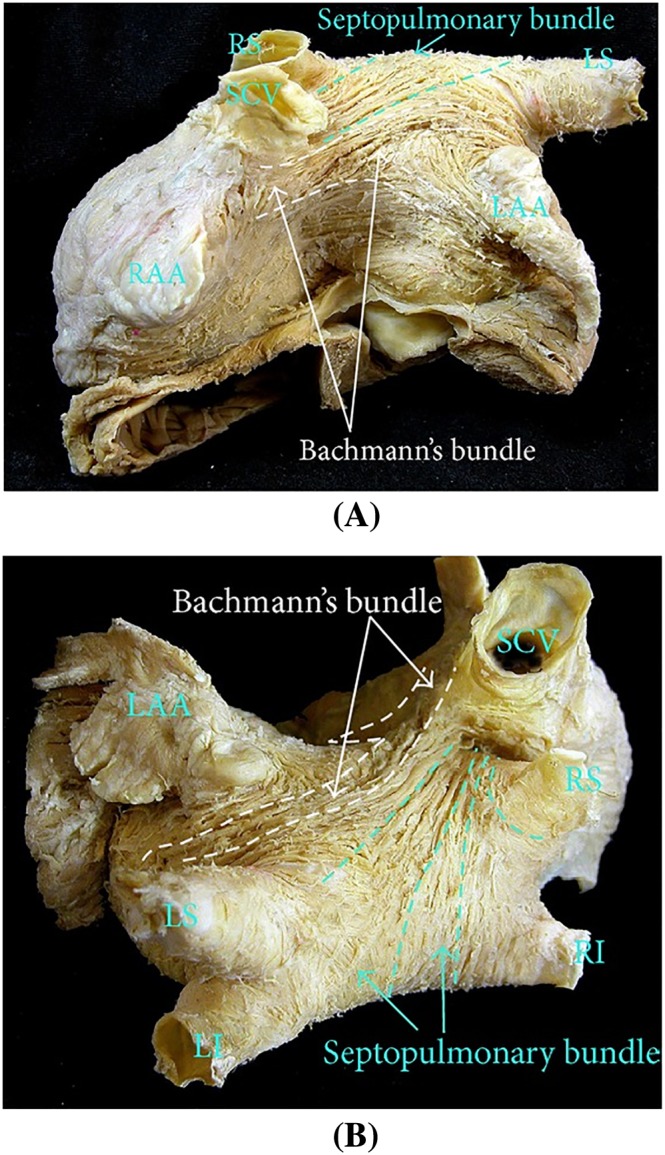
The left atrium fibre structure in the normal human heart. (A) Anterior view, where Bachmann's bundle runs from atrial septum to left atrial appendage, and the septopulmonary bundle runs obliquely to the superior wall. (B) Superior view where the septopulmonary bundle fans out. SCV, superior cava vein; RAA, right atrial appendage; LAA, left atrial appendage; LI, left inferior pulmonary vein; LS, left superior pulmonary vein; RI, right inferior pulmonary vein; RS, right superior pulmonary vein. The figure is from the work by Sánchez‐Quintana et al32