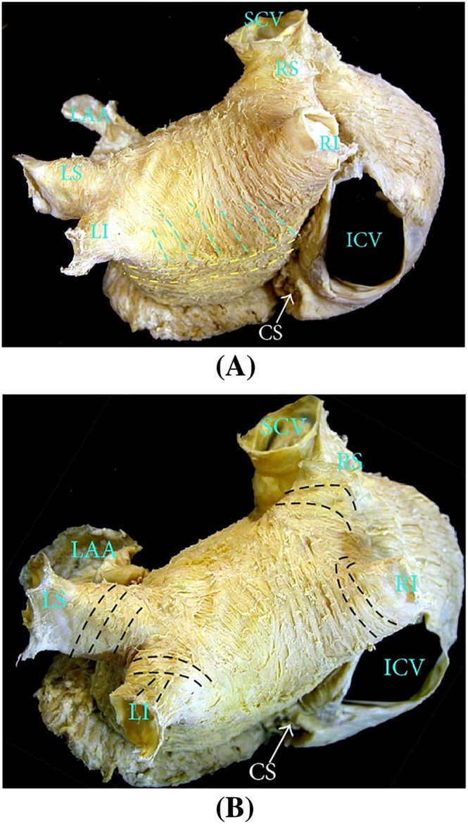
Figure A2.

The left atrium fibre structure in the normal human heart. (A) Posterior view, where the septopulmonary bundle meets the circumferential fibres coming from the lateral wall. (B) Near the pulmonary veins insertions where circular fibres are dominant and longitudinal, oblique fibres are also common. ICV, inferior cava vein; CS, coronary sinus. The figure is from the work by Sánchez‐Quintana et al32
