
Figure 3.
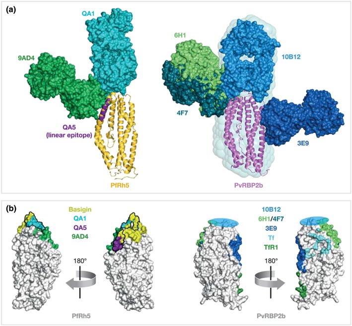
Epitopes of inhibitory mouse antibodies against PfRh5 and PvRBP2b. Structures of antigen‐Fab complexes are superimposed on the corresponding antigen. (a) Antigens PfRh5 (PDB ID 4WAT, yellow) and PvRBP2b (PDB ID 5W53, pink) are shown in ribbon representation, Fabs (each with heavy and light chains in the same colour) are in surface representation. The linear epitope of inhibitory antibody QA5 (PfRh5 residues 201–213) is indicated in purple. The SAXS‐derived model of the PvRBP2b‐10B12 complex is surrounded by its SAXS‐envelope. (b) Surface representation of PfRh5 and PvRBP2b (white). Antibody and receptor footprints are coloured with a 6 Å‐distance cut‐off. The rough binding region of 10B12 is indicated by a blue circle. Antibody epitopes of QA1 and QA5 directly overlap with the Basigin binding site of PfRh5