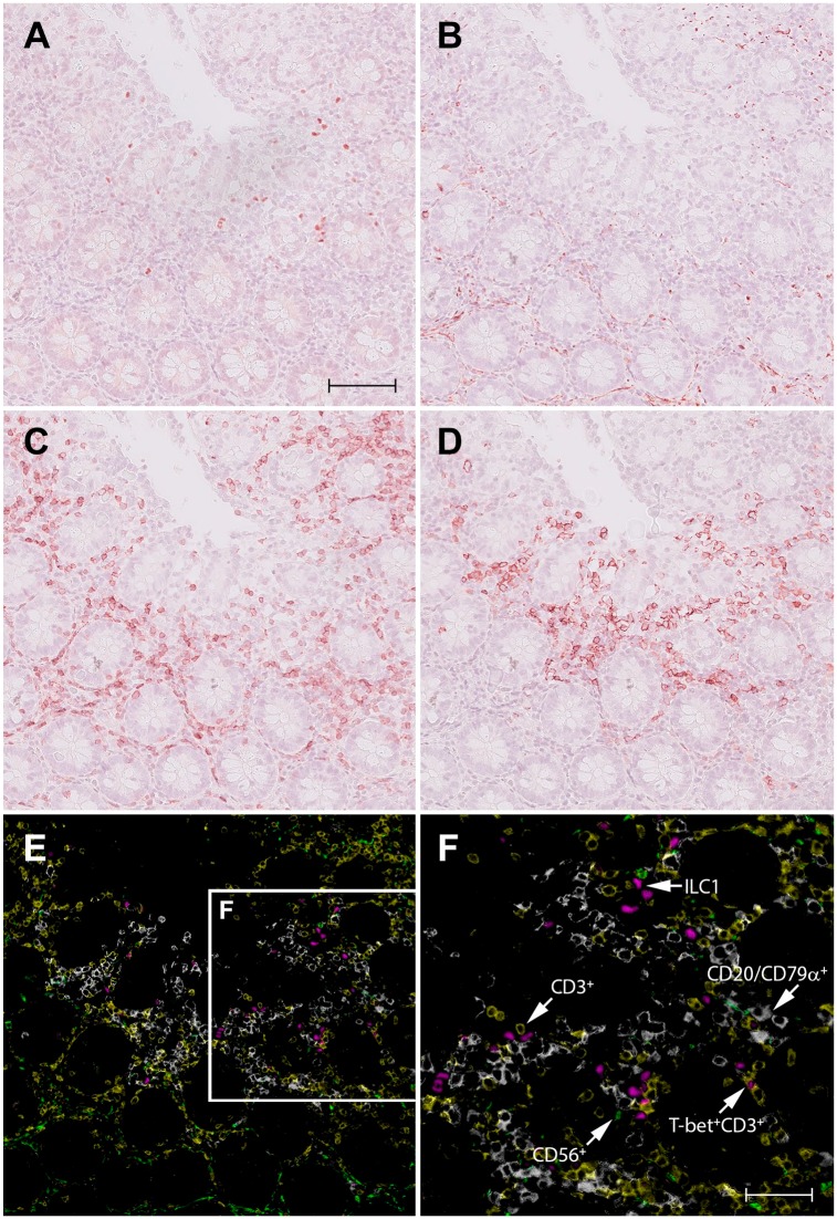
Figure 1.
Identification of ILC1s in normal human colon by virtual quadruple staining. A single section of colon tissue is successively stained with T-bet (A), CD56 (B), CD3 (C), and a mix of CD20/CD79α (D), using NovaRED as substrate and hematoxylin as counterstaining. Between each staining round, sections were digitized, decolorized, and stripped from antibodies. The red color in the digital images A–D, representing the immunopositive signal, is converted to and combined in one false color composite image (E): T-bet (magenta), CD56 (green), CD3 (yellow), and a mix of CD20/CD79α (white). Figure F shows a higher magnification of the framed area in (E). Scale bar in A (=B, C, D, E): 100 μm; F: 50 μm. Abbreviations: ILC, innate lymphoid cell.

