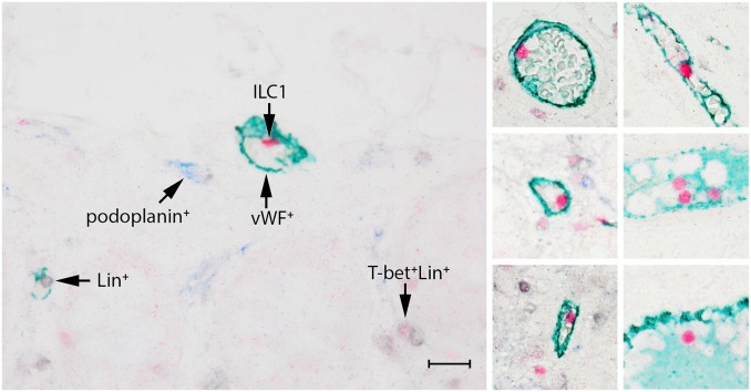
Figure 3.
ILC1s are frequently encountered in the lumen of blood vessels, but not in lymph vessels. ILC1s are identified by T-bet (red) and are Lin− (CD3−CD20/79α−, in black). Blood vessels are stained with vWF (green), lymph vessels with podoplanin (blue). Scale bar: 50 μm. Abbreviations: ILC, innate lymphoid cell.

