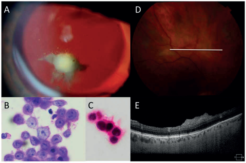
Figure 2:
Representative eyes 3 and 4: A: Slit lamp photography of the left eye demonstrating amelanotic vitreous opacities which were present in both eyes. B: Cytopathology of vitreous biopsy samples reveals atypical melanocytes harboring large irregularly bordered nuclei with prominent eosinophilic nucleoli. The attached pink cytoplasm also displays foci of melanin pigment deposition (haematoxylin eosin stain, 100X). C: Lesional cells reveal SOX-10 nuclear immunoreactivity (oil immersion, 100X) D: fundus photograph of right eye demonstrating amelanotic retinal infiltrates E: Optical coherence tomography of corresponding area demonstrating pre-retinal opacities.

