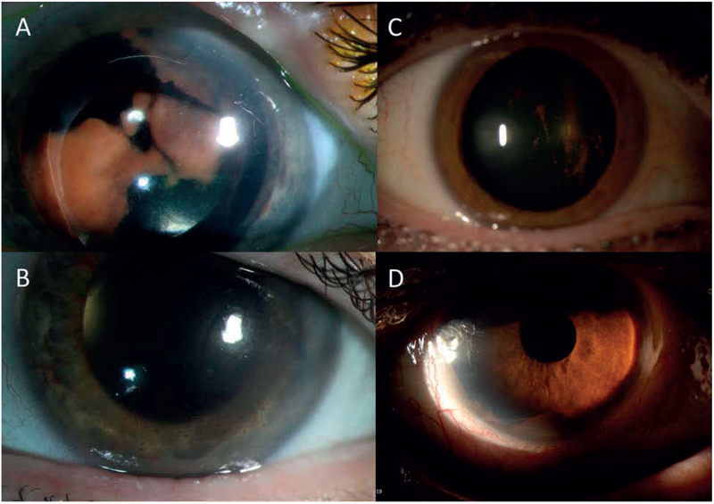
Figure 4:
Representative slit-lamp images of anterior chamber disease. A: Right eye (eye 2) demonstrating retrolental amelanotic/yellow sheet-like opacity. B: Response following 3 intravitreous melphalan injections and vitrectomy. C: Right eye (eye 7) on initial presentation demonstrating pigment deposition on the posterior surface of the crystalline lens and in the anterior vitreous D: Recurrence of disease featuring diffuse pigment clumping in the inferior angle of the anterior chamber.

