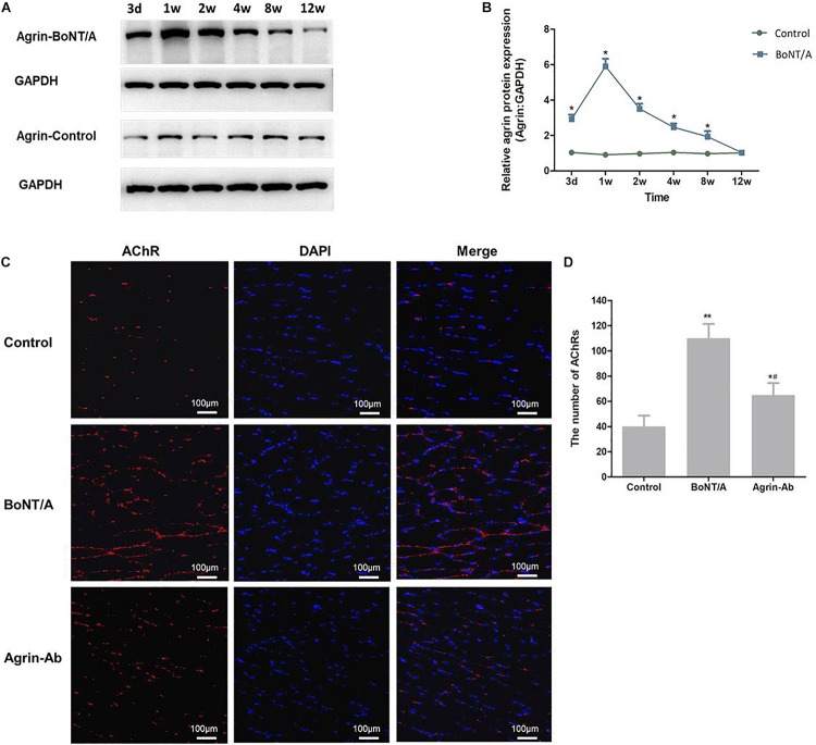
FIGURE 1.
Agrin participates in regulating nerve sprouting after BoNT/A application. (A) Western blot analysis of agrin at NMJs after BoNT/A injection. GAPDH was used as internal control. (B) Quantitation of agrin protein was performed using GeneTools from SynGene software and normalized to GAPDH. *P < 0.05 versus control. (C) α-Bungarotoxin staining analysis of the number of AChRs after BoNT/A and agrin-Ab injection at week 1. Nuclei were counterstained with DAPI (4′-6-diamidino-2-phenylindole; blue). Images were merged as indicated. Scale bar, 100 μm. (D) Number of AChRs; bars represent mean ± SD of three different experiments. *P < 0.05, **P < 0.01 compared to the control group. #P < 0.05 compared to the BoNT/A group.

