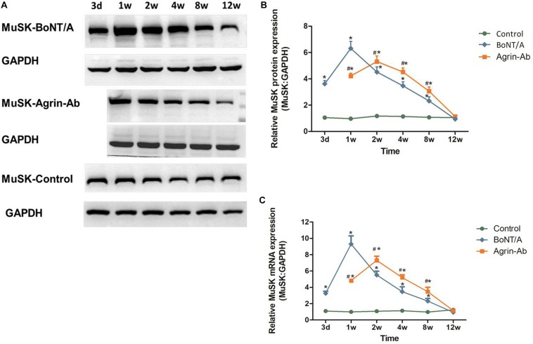
FIGURE 3.
Agrin-Ab delays the increase of MuSK expression. (A) Protein levels of MuSK were assayed by western blot after BoNT/A and agrin-Ab injection. GAPDH was used as an internal control. (B) Protein quantification was normalized to GAPDH and data are presented as mean ± SD of three different experiments. *P < 0.05 compared to the control group. #P < 0.05 compared to the BoNT/A group. (C) mRNA levels of MuSK were assayed by qPCR after BoNT/A and agrin-Ab injection. GAPDH was used as an internal control. Data represent the mean ± SD of three different experiments. *P < 0.05 compared to the control group. #P < 0.05 compared to the BoNT/A group.

