Figure 7.

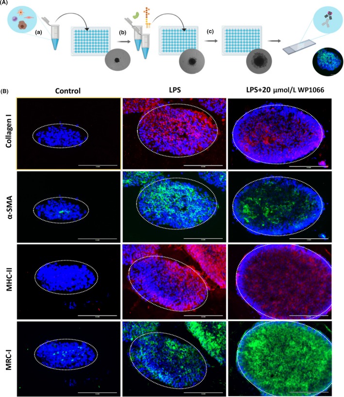
Therapeutic efficacy of WP1066 in human 3D spheroid model. A. Spheroid assay schematic illustration: (a) The cell mixture containing HepG2, LX2, HUVECS, and THP1 was plated in 96‐well ULA plates at a density of 1,200 cells/uL and grown for 7 d; (b) At day 7, spheroids were treated with or without LPS and WP1066; (c) After 7 d of treatment, the spheroids were retrieved, snap frozen, and sectioned. The corresponding cryosections were used for immunostaining. B. First panel of images depicts 3D spheroids stained with fibrosis marker, collagen I (in red); second panel depicts 3D spheroids stained with HSC activation marker, α‐SMA (in green); third panel of images presents 3D spheroids stained with macrophage activation marker, MHC‐II (in red); and fourth panel of images depicts 3D spheroids stained with M2 macrophage marker, mannose receptor (in green). Blue color shows DAPI nuclear staining. The data presented here represent n = 5 spheroids
