
Figure 2. Cryo-EM reconstruction of the EHEC T4P filament at ~8 Å resolution and PpdD pilin structure fitting.
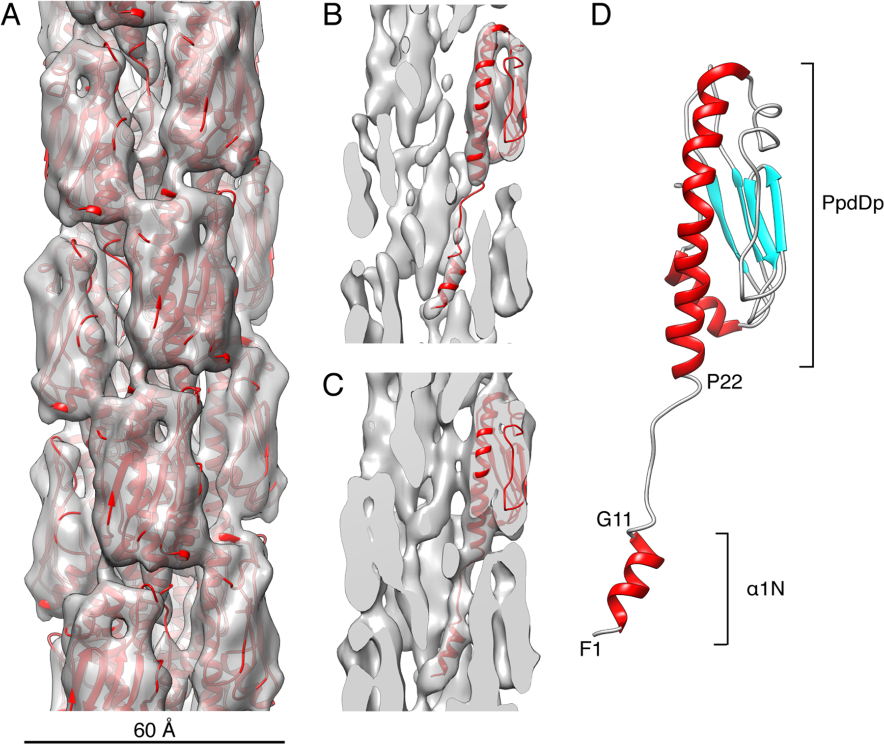
(A) Surface view of the EHEC T4P cryo-EM reconstruction at ~4.2σ contour level with the refined atomic model shown in ribbons (red). The diameter of the pilus is ~60Å. (B-C) Cross-sections of the cryo-EM reconstruction at ~5σ (B) and ~3.8σ (C) contour levels and with a single PpdD subunit shown in ribbons. (D) Structure of a PpdD pilin subunit in the refined EHEC T4P structure colored by secondary structures and showing the extended region between the short α1N helix and the PpdDp domain. The positions of helix breaking between residues G11 and P22 are indicated.