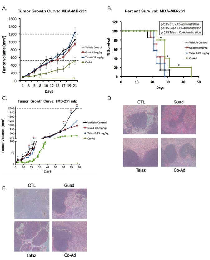
Figure 5. Co-administration of guadecitabine and talazoparib effective in TNBC xenograft model.
(A) MDA-MB-231 cells (2 × 10⁶) were subcutaneously injected into the flank of nude mice (total n=28) and then randomized to each group (n=7). When tumors reached 100mm3 mice were treated daily (five times per week) with guadecitabine (Guad; 0.5mg/kg, s.c.) or talazoparib (Talaz; 0.25mg/kg, orally) for three cycles. Tumor volume was measured at the indicated time points. Day 0 indicates treatment initiation. Statistically significant difference in tumor volume (until end of the study) is denoted by an asterisk and arrow. (B) Percent survival was measured by sacrifice of mice once tumor volume reached 2000mm3 or showed necrosis. The ‘#’ denotes mice which were removed due to formation of necrosis. (C) TMD-231 cells (0.5 × 10⁶) were injected into the mammary fat pad (mfp) of nude mice (total n=28) and then randomized to each group (n=7). When tumors reached 10mm3 mice were treated daily (five times per week) with Guad (0.5mg/kg, s.c.) or Talaz (0.25mg/kg, oral gavage) for three cycles. Tumor volume was measured at the indicated time points. Representative hematoxylin-eosin staining of (D) MDA-MB-231 tumors (E) TMD-231 tumors. * p<0.01, ** p<0.001, *** p<0.0001 compared to control.

