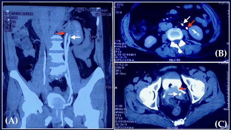
Figure 3. Post-operative computed tomography urogram.
(A) Coronal view showing the presence of two left ureters (red and white arrow). (B) Axial view showing orthotopic (red arrow) and ectopic (white arrow) left ureters in proximity to the left kidney. (C) Axial view at the level of bladder, showing the insertion of orthotopic ureter (red arrow) into the superior-lateral aspect. The transected ectopic ureter (white arrow) is not attached to the bladder.

