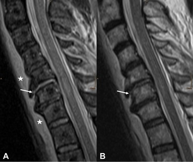
Figure 4.

A 70‐year‐old male patient after a heavy fall. Sagittal short tau inversion recovery (A) shows a clear interruption (white arrow) of the anterior longitudinal ligament (ALL) at C5/6. T2‐weighted images (B) do not show this interruption so clearly (white arrow), with poor demarcation of liquid accumulation in the area of interruption. Prevertebral hematoma can be depicted with both sequences with the maximum thickness in the segment directly adjacent to the pathological findings (asterisks in A).
