
Figure 2.
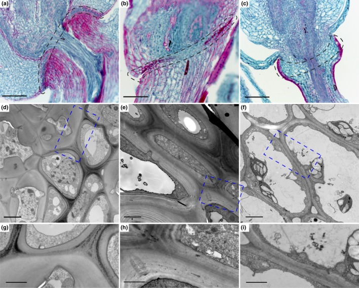
The abscission zones (AZ) of rice, Brachypodium and Setaria differ in histology and cell wall structures. (a–c) Fast green/Safranin O staining of mature spikelets in (a) rice, (b) Brachypodium and (c) Setaria. Safranin O stains secondary cell wall including lignin, suberin and cutin with a magenta colour, and fast green stains primary cell wall and cytoplasm with a green colour. The AZ is marked with black dotted lines. (d–f) Transmission electron microscopy images of the AZ and surrounding cells in (d) rice, (e) Brachypodium and (f) Setaria. (g–i) Zoomed in images of the blue dotted rectangles shown in (d‐f). Bars: (a ‐c) 100 µm; (d #x2010;f) 2 µm; (g #x2010;i) 1 µm.