Figure 1.

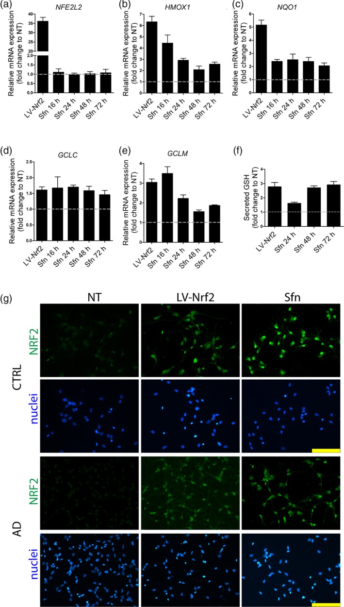
NRF2 induction in astrocytes by lentiviral transduction or Sfn treatment. (a) Relative mRNA expression of NRF2 (NFE2L2) after treatment shown as fold change to non‐treated (NT) astrocytes. Lentiviral transduction increases NRF2 expression, while Sfn treatment (5 μM) does not. (b–e) Relative mRNA expression of NRF2 target genes (HMOX1, NQO1, GCLC, and GCLM) after treatment. Both lentiviral transduction and Sfn treatment increase the expression of NRF2 target genes. (f) Glutathione secretion after treatment. Both lentiviral NRF2 and Sfn treatments increase the secretion of glutathione from astrocytes. (a–f) Experiments were performed with one isogenic pair and both AD and control cells behaved similarly. Representative data from one isogenic control line are shown as fold change to non‐treated astrocytes. Data are presented as mean ± SEM from three independent experiments. (g) Representative immunofluorescence images of NRF2 (green) staining show the accumulation of NRF2 in the nucleus upon both treatments in astrocytes of both genotypes. Nuclei were counterstained with Hoechst 33258 (blue). Scale bars 100 μm. (NT: non‐treated cells; GSH: glutathione; LV‐NRF2: lentiviral NRF2 transduction; Sfn: sulforaphane; CTRL: isogenic control astrocytes; AD: PSEN1 ΔE9 mutated Alzheimer's disease astrocytes) [Color figure can be viewed at http://wileyonlinelibrary.com]
