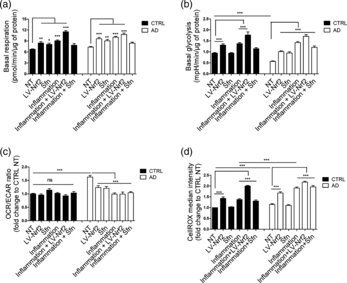
Figure 2.

Both inflammatory stimulation and NRF2 induction lead to metabolic activation of astrocytes. (a) Quantification of the basal respiration rates from the oxygen consumption rate analyzed by Seahorse XF analyzer in the presence of glucose. Both inflammatory stimulation and NRF2 induction activate oxidative metabolism in both cell types. (b) Quantification of the basal glycolytic activity from extracellular acidification rate. Both inflammatory stimulation and LV‐NRF2 induction activate glycolytic metabolism in both cell types. (c) Relative OCR/ECAR ratio after glucose injection shown as fold change to non‐treated isogenic control astrocytes. Both inflammation and NRF2 induction reduce the ratio of oxidative to glycolytic metabolism in AD cells. (a–c) All data are presented as mean ± SEM, n = 4–6 independent experiments using two isogenic pairs with 3–4 technical replicates in each experiment. (d) Reactive oxygen species (ROS) production, measured by CellROX fluorogenic probe and FACS analysis. As expected, the increased respiratory activity upon inflammatory stimulation or NRF2 induction also leads to increased ROS production in both AD and isogenic control cells. However, in Sfn‐treated cells, ROS is not increased in either genotype. Relative results compared with non‐treated control cells shown as mean ± SEM, n = 3 independent experiments with two isogenic pairs. ***p < .001; **p < .01; *p < .05. ns, non‐significant
