Figure 3.

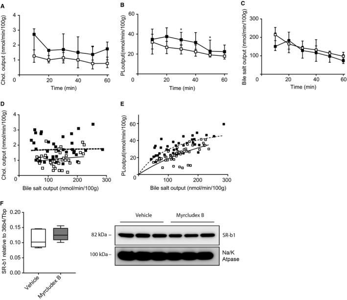
Increased biliary cholesterol and phospholipid secretion is not mediated by SR‐B1. (A) Cholesterol output, (B) phospholipid output, and (C) bile salt output into bile (in nmol/min/100 g BW) in Sr‐bI −/− mice. (D) Cholesterol output and (E) phospholipid output were plotted as a function of biliary bile salt (linear regression was performed on log‐transformed data and significance was assessed by comparing slopes or intercepts). (F) Hepatic mRNA and protein expression level of Sr‐bI in WT mice. RNA expression is compared to the geometric mean of reference genes 36b4 and Tbp. Protein levels were compared to the sodium/potassium ATPase (n = 3/group). Data are presented as median and interquartile range. White squares/bars indicate the vehicle group, and black/gray squares/bars indicate the Myrcludex B group. Differences between groups were analyzed using the Mann‐Whitney U test. Asterisk (“*”) indicates P < 0.05; n = 8 mice/group. Abbreviations: Chol., cholesterol; PL, phospholipid; Tbp, TATA‐box binding protein.
