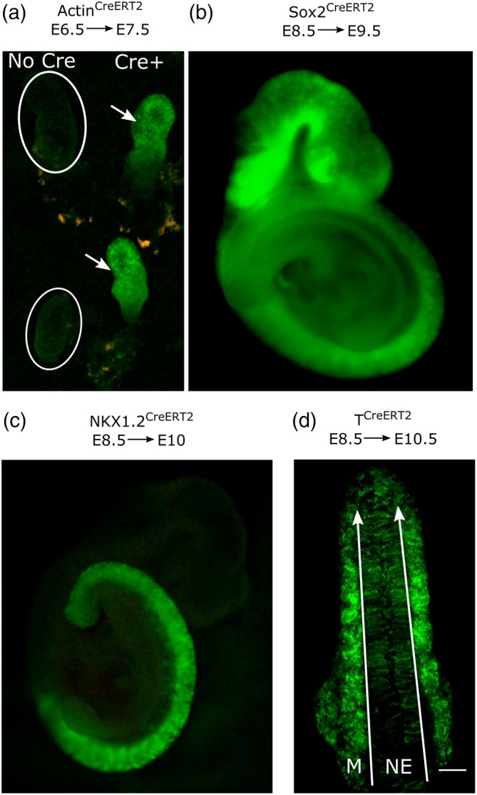
Figure 1.

Pipette feeding of tamoxifen to pregnant female mice activates various embryonic CreERT2 drivers relevant for developmental biology research. (a–d) CreERT‐negative female mice were lightly scruffed and fed a single dose of 75 μl peanut oil containing 100 mg/ml tamoxifen free base. Recombination of Rosa26 YFP (b) or Rosa26 mTmG (a, c, d) reporters, producing green fluorescence. (a) Stereoscope image of Actin CreERT2‐positive (arrows) and negative (circles) embryos collected at E7.5, 24 h after tamoxifen administration. (b) Stereoscope image showing extensive Sox2 CreERT2 recombination in an embryo collected at E8.5, 24 h after tamoxifen. (c) Stereoscope image showing Nkx1.2 CreERT2 recombination in an embryo collected at E10, 36 h after tamoxifen. (d) Confocal image of a dorsal view of the caudal end of a T CreERT2–expressing embryo showing lineage tracing of both neuroepithelial (NE) and mesodermal (M) populations. Long arrows indicate the tail end in the confocal image; scale bar = 100 μm
