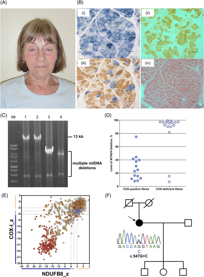
Figure 1.

Clinical, histopathologic and molecular characterisation of a patient harbouring a novel heterozygous c.547G>C GMPR variant. A, Ophthalmological features of the patient with PEO harbouring a novel heterozygous GMPR variant, highlighting bilateral ptosis and frontalis muscle hyperactivity. B, A skeletal muscle biopsy from the patient was subjected to (a) COX, (b) SDH, (c) sequential COX‐SDH histochemical reactions and (d) haemotoxylin and eosin (H&E) staining. Scale bar represents 100 μM. C, 13‐kb long‐range PCR assay of skeletal muscle mtDNA demonstrating multiple mtDNA deletions in the patient (lane 4) compared with aged‐matched controls (lanes 1 and 2) and a patient with a single, large‐scale mtDNA deletion (lane 3). D, Quantitative single‐fibre real‐time PCR assay reveals that the majority of COX‐deficient fibres exhibit clonally expanded multiple mtDNA deletions involving the MTND4 gene. E, Mitochondrial respiratory chain expression profile showing NDUFB8 (complex I), COX‐I (complex IV) and porin levels in individual patient skeletal muscle fibres. Each dot represents an individual muscle fibre, colour coded according to mitochondrial mass (very low, blue; low, light blue; normal, light orange; high, orange; and very high, red). Black dashed lines represent the SD limits for the classification of fibres. Lines adjacent to the X‐ and Y‐axis represent the levels of NDUFB8 and COX‐I (beige, normal; light beige, intermediate (+); light blue, intermediate (−); and blue, negative]. F, Family pedigree and Sanger sequencing confirmation of the novel c.547G>C GMPR variant in the index case [Colour figure can be viewed at http://wileyonlinelibrary.com]
