
Figure 2.
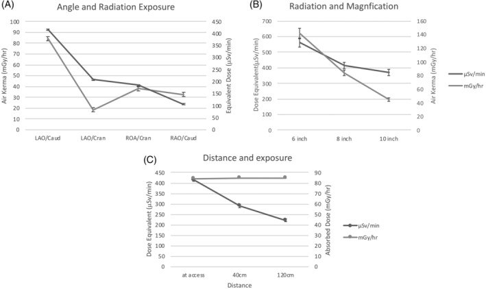
(a) Comparison of radiation exposure to the mannequin and human computational phantom with four angles of the detector. Left anterior oblique (LAO); Right anterior oblique (RAO); caudal (Caud); cranial (Cran). LAO/Caud with significantly more radiation measured by μSv/min and mGy/hr, p < .005. (b) Increase in magnification significantly increases exposure measured with μSv/min and mGy/hr (p < .005). (c) Significant decline in μSv/min (p < .001) with no change in mGy/hr produced by the X‐ray tube (p = .732) when mannequin positioned 40 and 120 cm from femoral access site of phantom