
Figure 1.
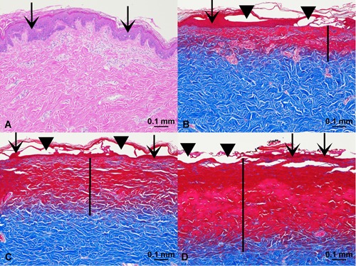
Acute tissue injury. (A) Control, hematoxylin and eosin (H&E) stain, ×10 magnification. (B) Helium plasma 20% power (single pass), Masson's trichrome stain, ×10 magnification. (C) Helium plasma 40% power (single pass), Masson's trichrome stain, ×10 magnification. (D) Nitrogen plasma PSR3 (4.0 J), Masson's trichrome stain, ×10 magnification. Scale bars = 0.1 mm. Vertical black bars designate depth of dermal injury, extending into the papillary and reticular dermis. Scarlet stained tissue in 1B, C, D indicates denatured tissue characterized by coagulative necrosis.