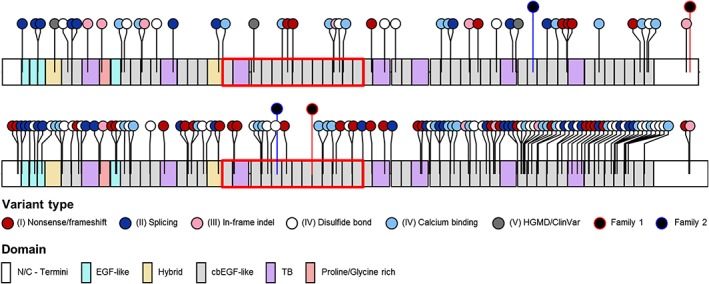
Figure 2.

FBN1 and FBN2 a priori (likely) pathogenic variants in gnomAD and FBN1/FBN2 dual variants in Family 1 and Family 2. Lollipops show the type and position of variants in relation to protein domain structure. Red boxes indicate the severe/neonatal region in FBN1 (exons 23‐34) and the comparable congenital contractural arachnodactyly (CCA)‐mutation‐hotspot region in FBN2 (exons 23‐34). cbEGF, calcium‐binding epidermal growth factor; EGF, epidermal growth factor; HGMD, Human Gene Mutation Database; indel, small insertion/deletion; TB, transforming growth factor β binding. Information on protein domains was obtained from umd.be/FBN1 and umd.be/FBN2. Lollipop diagrams were generated using the R package “trackViewer,” available from http://bioconductor.org/packages/release/bioc/html/trackViewer.html [Colour figure can be viewed at http://wileyonlinelibrary.com]
