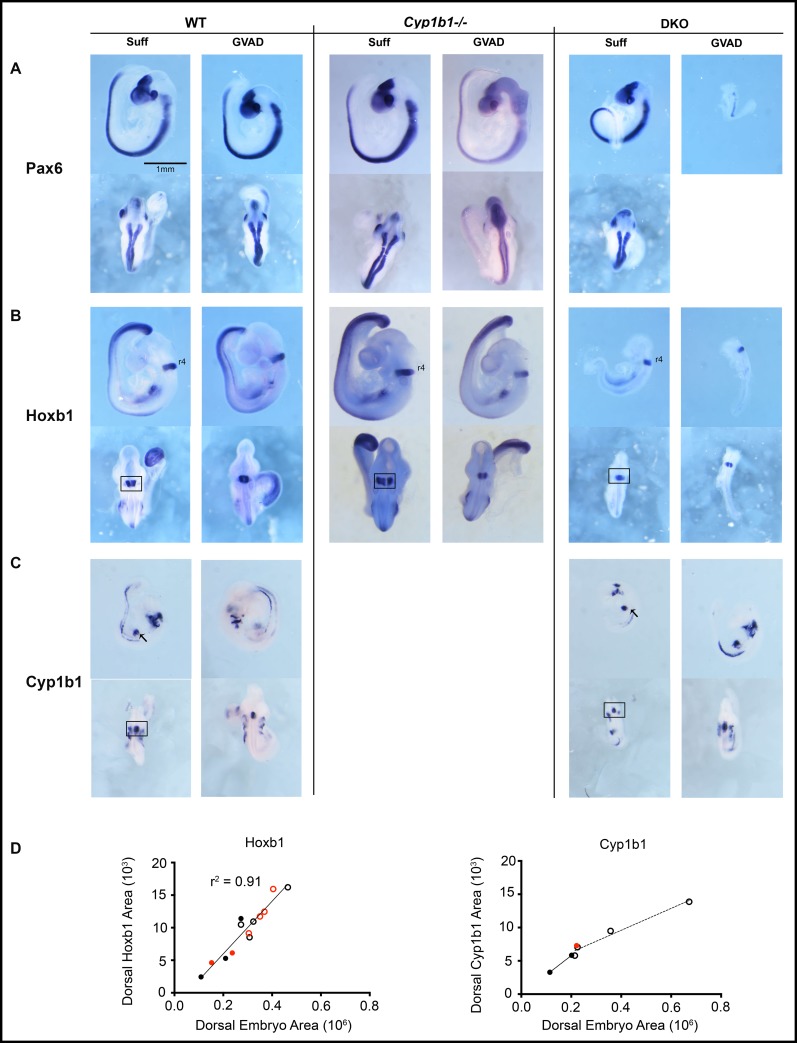
Fig 7. Effects of GVAD on morphogenic gene expression patterns in WT, Cyp1b1-/-, and DKO embryos.
E9.5 WT (left), Cyp1b1-/- (middle), and DKO (right) embryos from dams administered vitamin A-sufficient (Suff) or -deficient (GVAD) diets are shown. DKO embryos are compared to WT embryos in a similar size range (Fig 2A). Embryos were probed by ISH to evaluate expression patterns of Pax6 (A), Hoxb1 (B), and Cyp1b1 (C). Lateral (upper) and dorsal (lower) views are provided. GVAD DKO embryos were very variable in size. A third were similar to Suff DKO, while the remainder were smaller and often malformed. All images are shown to the same scale. Analysis of r4 absolute expression areas of Hoxb1 and Cyp1b1 each from a dorsal view of ISH images from WT (open circles) and DKO embryos (solid circles) from Suff (black) or GVAD (red) diets (D). For ISH images see S4 Fig.

