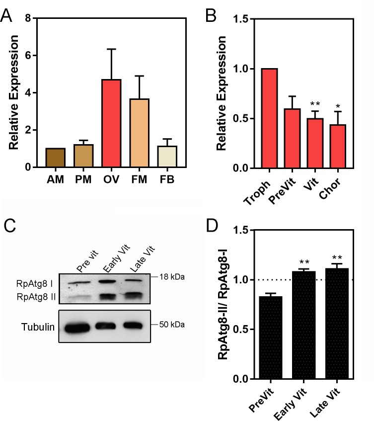
Fig 1. RpATG8 is highly expressed in the ovaries of vitellogenic females and autophagosomes are formed during vitellogenesis in the oocytes.
A. RpATG8 mRNA quantification in the different organs of vitellogenic females dissected 7 days after the blood meal. B. RpATG8 mRNA quantification in the different components of the ovariole: Tropharium, pre-vitellogenic oocytes, vitellogenic oocytes and chorionated oocytes. The relative expression was quantified using the ΔΔCT method. Graphs show mean ± SEM (n = 6). C. Immunoblotting using the antibodies raised against RpATG8 (LC3). Lanes 1–3: Pre vitellogenic, early vitellogenic and late vitellogenic oocytes. RpATG8-II: RpATG8 conjugated to the phosphatidylethanolamine. D. Immunoblotting densitometry showing the ratio of RpATG8-II/RpATG8-I (n = 3). *p<0.05, **p<0.01. One Way ANOVA.

