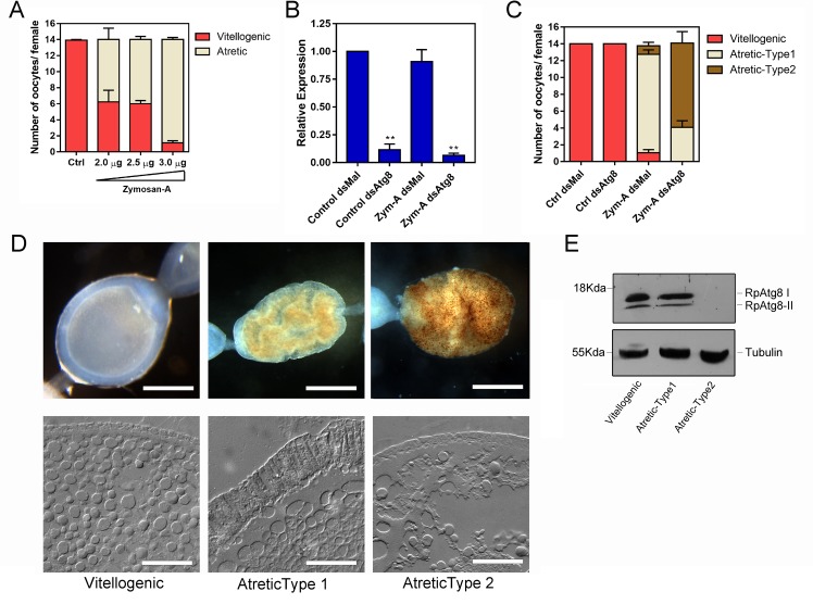
Fig 3. Silencing of RpATG8 results in the same number of atretic oocytes, but with a different morphology.
A. Increasing concentrations of Zymosan-A were directly injected in the hemocoel of 10 vitellogenic females per treatment 3 days after the blood meal and the number of atretic oocytes was accessed 7 days after the blood meal. Graph shows mean ± SEM (n = 10). B. Levels of RpATG8 mRNA silencing in control and Zymosan-A-challenged females, 7 days after the blood meal. dsMal: control dsRNA, dsATG8: dsRNA designed to specifically target the RpATG8 sequence. Graph shows mean ± SEM (n = 6). **p<0.01, ***p<0.001, One Way ANOVA. C. The challenge with Zymosan-A was performed in control and silenced females and the number and types of atretic oocytes were accessed in 10 insects per treatment. Graph shows mean ± SEM (n = 10). D. Both types of atretic oocytes were observed under the stereomicroscope (Upper panel, Bars: 0.5 mm) and cryosections were observed in the light microscope operating in differential interferential contrast mode (Lower panel). Bars: 500 μm. E. Immunoblotting to test the silencing of RpATG8 in control and atretic oocytes. Tubulin was used as loading control (n = 3).

