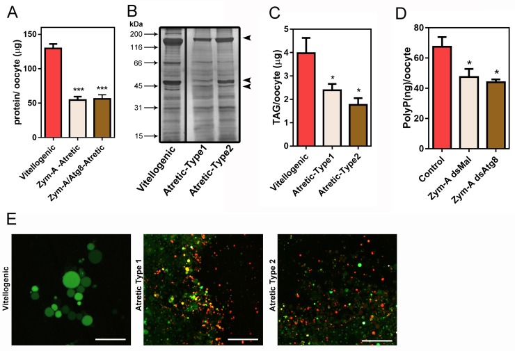
Fig 4. Silencing of RpATG8 does not affect degradation of the main yolk macromolecules during follicular atresia.
A. Total protein quantifications in vitellogenic, control atretic (Type1) and silenced atretic (Type2) oocytes (n = 7). B. 10% SDS-PAGE showing the protein profile of vitellogenic and both types of atretic oocytes (n = 3). Arrows indicate vitellogenin subunits. C. TAG content detected in vitellogenic and both types of atretic oocytes (n = 6). D. PolyP content detected in vitellogenic and both types of atretic oocytes (n = 4). E. The yolk organelles from each of the oocytes (vitellogenic, atretic type-1 and atretic type-2) were incubated with 5μg/ml Acridine Orange (AO) and observed under the fluorescence microscope (n = 5). Bars: 50 μm. *p<0.05, ***p<0.001, One Way ANOVA.

