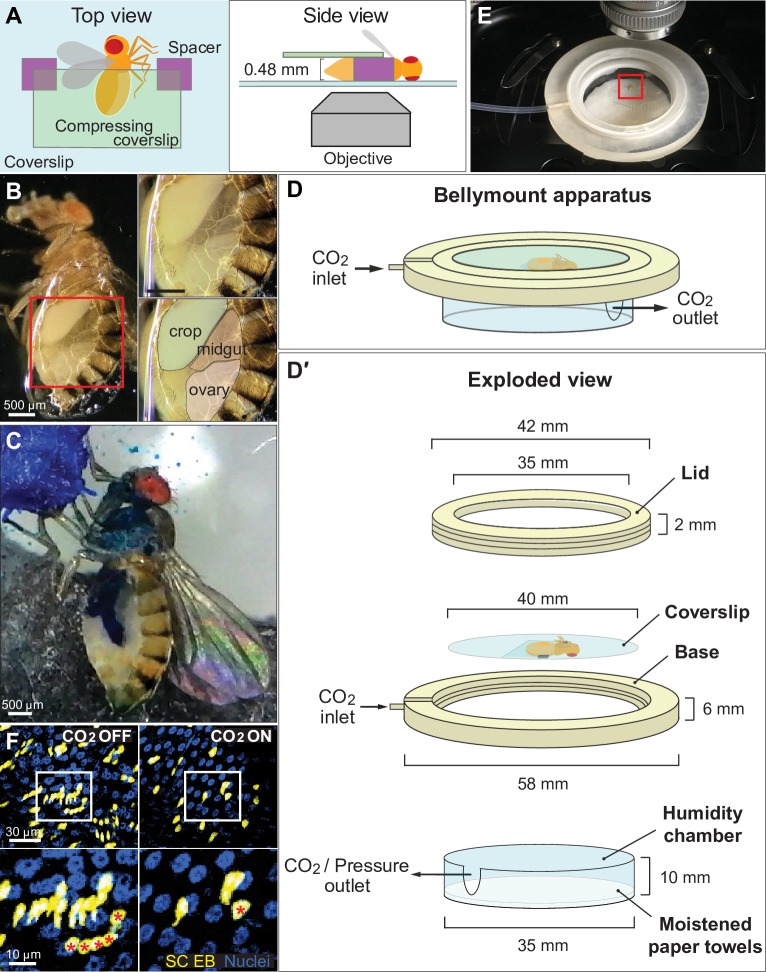
Fig 1. The Bellymount platform enables intravital imaging of the adult Drosophila abdomen.
(A) Cartoons of Bellymounted animal. Top and side views are shown. The cuticle of the ventrolateral abdomen is glued to the bottom coverslip. To maximize contact between the cuticle and the glue, a second, smaller coverslip gently compresses the abdomen. (B) Underside view of a Bellymounted adult female. Gluing caused the ventral cuticle to become light transparent. The edge of the glue patch appeared as a refractive line around the abdomen. Right panels are close-ups of boxed region in left panel. (C) Bellymounted animals ingest nutrients, undergo GI transit, and defecate. As a mounted animal ingested blue-colored sugar water, its midgut gradually became colored blue. Image shows a single time point from S3 Movie. (D) Cartoon of Bellymount apparatus for delivery of CO2 anesthesia during confocal imaging. Isometric drawing shows assembled (D) and exploded (D′) configurations. The coverslip with the glued animal sits inside the base of the apparatus. CO2 flows through the indicated ports. CAD files for 3D printing of the lid and base are provided as S1 and S2 Files. See also S2 Fig. (E) Bellymount apparatus positioned for imaging on an upright microscope. Red box shows the location of the glued animal. (F) CO2 anesthesia minimizes tissue movements during acquisition of confocal z-stacks. Without CO2 (left), tissue movements during z-stack acquisition caused individual cells to be represented multiple times. With CO2 (right), tissue movements were inhibited, and z-stack images were accurate. Bottom panels are close-ups of boxed regions in top panels. Red asterisks label the identical cell in images taken without and with CO2. Blue, nuclei; yellow, midgut stem cells and enteroblasts. Images are projections of confocal stacks. Genotype: esg>LifeActGFP; ubi-his2av::mRFP. See S4 Movie. CAD, computer-aided design; EB, enteroblast; esg, escargot-Gal4; GFP, green fluorescent protein; GI, gastrointestinal; his2av, histone variant His2av; mRFP, monomeric red fluorescent protein; RFP, red fluorescent protein; SC, stem cell.

