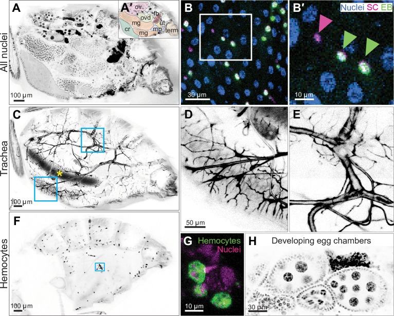
Fig 2. Whole-abdomen, micron-resolution imaging of organs, tissues, and cells in live, intact animals.
(A) Native arrangement of abdominal organs. Visible organs include the crop (cr), midgut (mg), ovary (ov), oviduct (ovd), fat body (fb), uterus (ut), Malphigian tubules (mp), and terminalia (term). Inverted grayscale, nuclei. Genotype: esg>his2b::CFP, GBE-Su(H)-GFP::nls; ubi-his2av::mRFP (only RFP is shown). See S5 Movie. (B) Nuclear morphologies of individual midgut cells. Immature diploid cells (SCs [magenta nuclei] and terminal EBs [green nuclei]) were dispersed among mature, polyploid enterocytes (blue nuclei). Right panel is a close-up of boxed region in left panel. Arrowheads indicate nuclei of a stem cell (magenta) and two enteroblasts (green). Genotype: esg>his2b::CFP, GBE-Su(H)-GFP::nls; ubi-his2av::mRFP. (C) The abdominal tracheal network. Whole-abdomen imaging showed connectivity of tracheal branches from cuticular spiracles to internal organs. Ingested food in the midgut lumen exhibited autofluorescence (yellow asterisk). Boxed areas are shown as close-ups in D and E. (D) An extensive network of secondary and tertiary trachea wrap around the midgut tube. (E) Primary trachea exhibit branching near their origin at the cuticular spiracle. Genotype in C–E: breathless>GFP; ubi-his2av::mRFP (only GFP is shown). (F) Whole-abdomen distribution of hemocytes. Individual hemocytes tended to localize to pigmented regions of abdominal tergites. (G) Morphology of individual hemocytes. Image is a close-up of boxed region in F. Scale bar, 30 μm. Genotype in F–G: hml>GFP; ubi-his2av::mRFP (only the GFP channel is shown in F). (H) Developing egg chambers. Each chamber included the immature oocyte and polyploid nurse cells that are surrounded by diploid follicle cells. Four chambers, arranged by developmental stage, are aligned within one ovariole at the bottom. One chamber from a different ovariole is at the top. Nuclei, inverted grayscale. Genotype: ubi-his2av::mRFP. See S6 Movie. All images are projections of confocal stacks. CFP, cyan fluorescent protein; EB, enteroblast; esg, escargot-Gal4; GBE, Grainyhead binding element; GFP, green fluorescent protein; hml, hemolectin; his2av, histone variant His2av; mRFP, monomeric red fluorescent protein; nls, nuclear localization sequence; RFP, red fluorescent protein; SC, stem cell; Su(H), Suppressor of Hairless.

