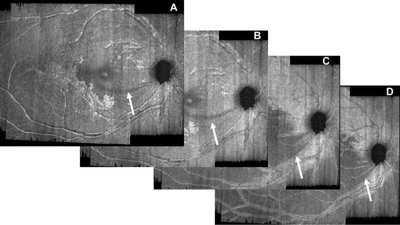
FIGURE 3.

A selection of images to illustrate scrolling through the retinal nerve fiber layer: at 8 μm (A), at 16 μm (B), at 36 μm (C), and at 54 μm (D). An arcuate defect inferior-temporal is seen (white arrow) in panel A and continues to develop through to panel D.
