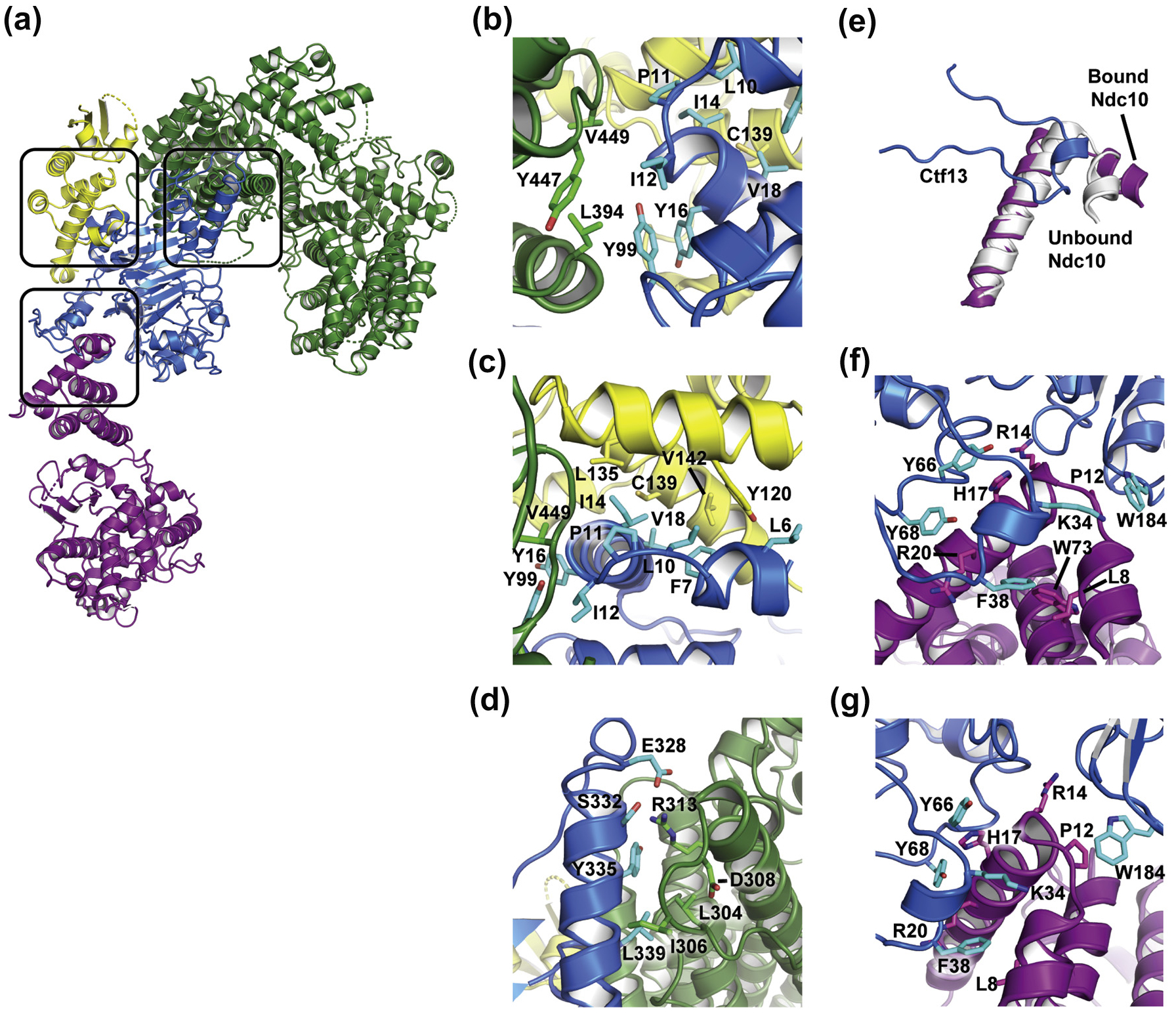
Fig. 3.

Interactions between subunits of the K. lactis CBF3–Ndc10 D1D2 complex, (a) Overall structure with Ctf 13–Skp1–Cep3b (F-box) interface, separate Ctf13–Cep3b interface, and Ctf13–Ndc10 D1D2 interface highlighted in black boxes, (b–c) Two views of residues at the F-box interface, (d) The Ctf13–Cep3b interface, (e) Isolated view of Ctf 13 residues 28–48 (blue) with the first two helices of Ndc10 D1D2 from the complex (purple) and from the published crystal structure (PDB 3SQI, gray), showing a reorientation of the first, shorter helix to accommodate interactions with Ctf13. (f–g) Two views of residues at the interface between Ctf13 and Ndc10 D1D2.
