Figure 2.

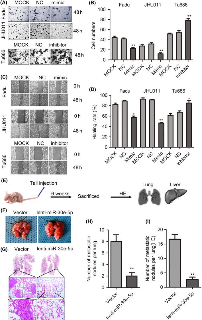
miR‐30e‐5p impedes squamous cell carcinoma of head and neck (SCCHN) metastasis. A and B, Cell invasion was assessed using a transwell assay. Representative fields with migrated cells (A) and quantification of the number of migration cells (B) are shown in the insets. C and D, The scratch test was used to check the changes in healing ability in vitro (C) and the percentage of healing area was determined (D). “The healing rate” is the ratio of “healing area” to “area of original wound.” E, Distant lung metastatic model was established via the tail vein injection of Fadu cells that stably overexpressed miR‐30e‐5p or vector. F and H, The images of macroscopic lung metastatic nodules were photographed (F), and numbers of lung nodules were calculated (H); arrowheads indicate metastatic nodules. G and I, Representative images (G) and quantification (I) of microscopic lung metastatic nodules stained with H&E. *P < 0.05; **P < 0.01
