Figure 3.

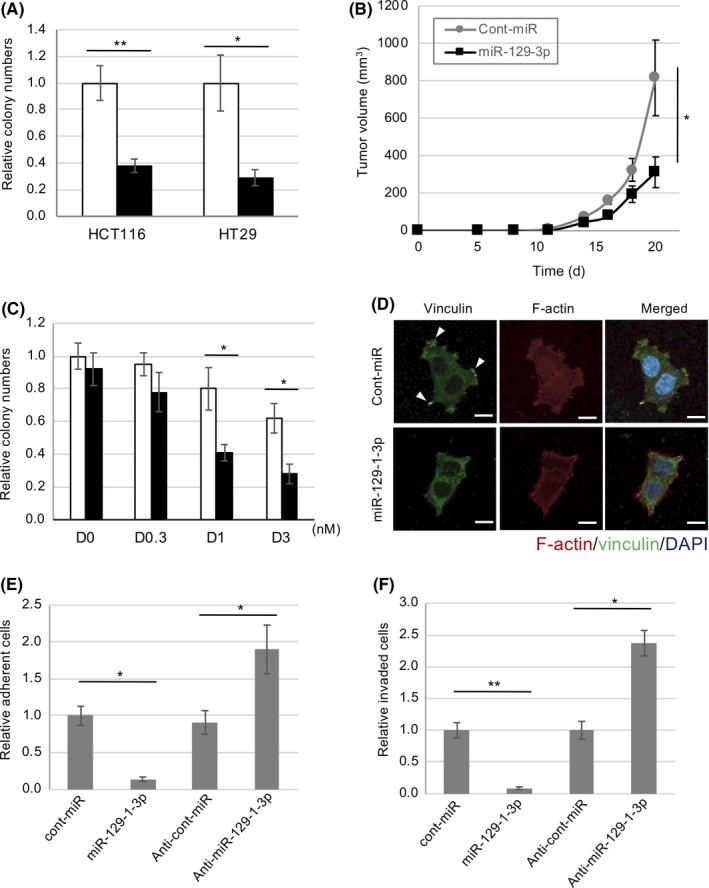
MicroRNA (miR)‐129‐1‐3p as a suppressor of tumor growth and malignancy in human colon cancer cells. A, HT29 and HCT116 cells were transfected with miR‐129‐1‐3p (black bar) or control‐miR (white bar) and subjected to soft‐agar colony formation assays. Relative colony numbers ± SD were obtained from 3 independent experiments. B, HT29 cells transfected with or without miR‐129‐1‐3p were inoculated s.c. into nude mice. Means ± SD of tumor volumes (mm3) obtained from 5 mice are plotted vs. time after inoculation (d). C, miR‐129‐1‐3p increased the sensitivity of HT29 cell growth to dasatinib (D). HT29 cells transfected with control (white bar) or miR‐129‐1‐3p (black bar) were treated with dasatinib at different concentrations, and cell growth was analyzed 3 days after dasatinib treatment. D, HCT116 cells were transfected with miR‐129‐1‐3p or control‐miR, and then subjected to immunocytochemistry. Actin filaments (red), vinculin, a marker of focal contact (green), and DAPI (blue) were analyzed by immunostaining HCT116 cells grown on fibronectin‐coated dishes. Scale bar = 20 µm. E, Cell adhesion assay on fibronectin of HCT116 cells treated with control, miR‐129‐1‐3p, or anti‐miR‐129‐1‐3p. F, In vitro invasiveness of the HCT116 cells used in (E). After 48 h, membranes were detached and cells were stained and counted. Relative number of cells per mm2 ± SD was obtained from 3 independent experiments (A–C, E, F). *P < .05, **P < .01 by Student’s t test
