
Figure 2.
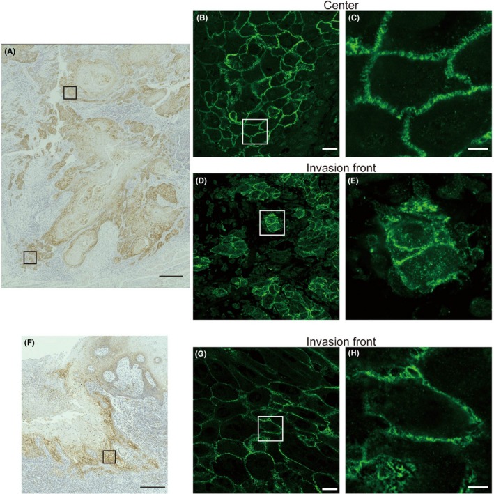
Typical cases of tongue squamous cell carcinoma (TSCC) showing either intracellular (A‐E) or membrane‐confined localization (F‐H) of claudin‐1 at the invasion front. Immunohistochemical (A, F) and immunofluorescence (B‐E, G, H) staining of claudin‐1 in two cases of TSCC. Upper and lower boxed areas in (A) show the center and the invasion front of the lesion, which are magnified in (B) and (D), respectively. The boxed area in (F) shows the invasion front of the lesion in another TSCC, which is magnified in (G). The boxed areas in (B, D, G) are further magnified in (C, E, H), respectively. See also Videos [Link], [Link], [Link], which correspond to (C, E, H), respectively. Scale bars: 300 μm (A, F), 25 μm (B, D, G), and 5 μm (C, E, H)