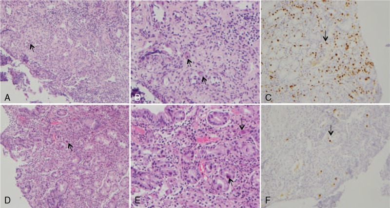
Figure 3.

Histopathology of cytomegalovirus (CMV) gastritis in Patient 3. (A) Histological detection of CMV inclusion bodies (arrow), biopsy specimen of a gastric ulcer. Hematoxylin and eosin. ×100. (B) Histological detection of CMV inclusion bodies (arrow), biopsy specimen of a gastric ulcer. Hematoxylin and eosin. ×200. (C) Positive CMV immunohistochemistry (arrow). Biopsy specimen of a gastric ulcer. × 100. (D) Histological detection of CMV inclusion bodies (arrow), biopsy specimen of an esophageal ulcer. Hematoxylin and eosin. × 100. (E) Histological detection of CMV inclusion bodies (arrow), biopsy specimen of an esophageal ulcer. Hematoxylin and eosin. ×200. (F) Positive CMV immunohistochemistry (arrow), biopsy specimen of an esophageal ulcer. ×100.
