
Figure 3.
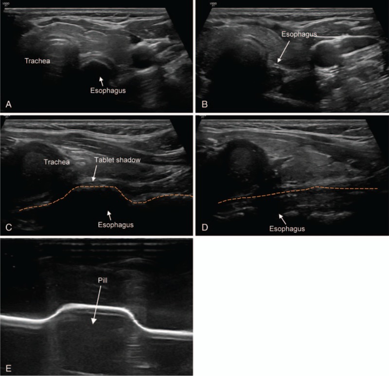
A 32-year-old woman (case 2) with discomfort on neck that occurred 2 h after taking a pill. The white arrow in each figure indicates the structure such as trachea and esophagus. The name of the structures is written at the end of the arrow or above the structure. (A) Axial view showing bulging esophagus due to impaction of pill. (B) Axial view showing collapsed esophagus after the pill went down. (C) Longitudinal view showing focal bulging esophagus due to impaction of pill. Dash line outlines the esophagus. (D) Longitudinal view showing collapsed esophagus after the pill went down. Dash line outlines the esophagus. (E) Simulated sonographic image of a pill. After the latex glove was filled with water, a pill was placed under the glove and the probe was placed on the glove to obtain a sonographic image of the pill. The pill was shown as hypoechoic lesion in the ultrasound.