Figure 1.

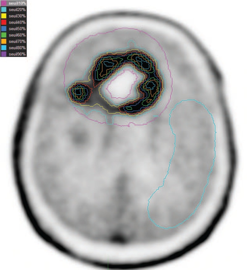
Various isocontours (from 10% to 90%) located on glioma and a 2nd region of reference (background activity) in an area of normal brain tissue including white and gray matter have been illustrated.

Various isocontours (from 10% to 90%) located on glioma and a 2nd region of reference (background activity) in an area of normal brain tissue including white and gray matter have been illustrated.