
Figure 3.
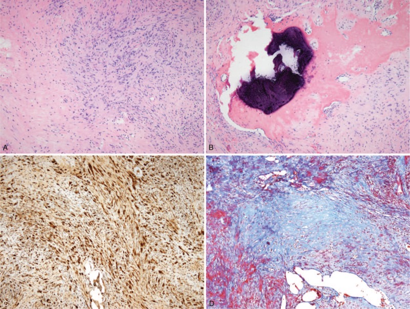
Histopathological examination of the surgical specimen. (A) Histopathological examination of the surgical specimen revealed a schwannoma, composed of compact areas of spindle cells (Antoni A, arrowheads) and loosely arranged foci (Antoni B, asterisk). (B) A small calcification and ossification (arrow) were observed. (C) Immunohistochemistry shows positive staining for the S-100 protein, confirming the diagnosis of schwannoma. (D) In this Masson trichrome-stained section, collagen deposition was revealed as blue areas.