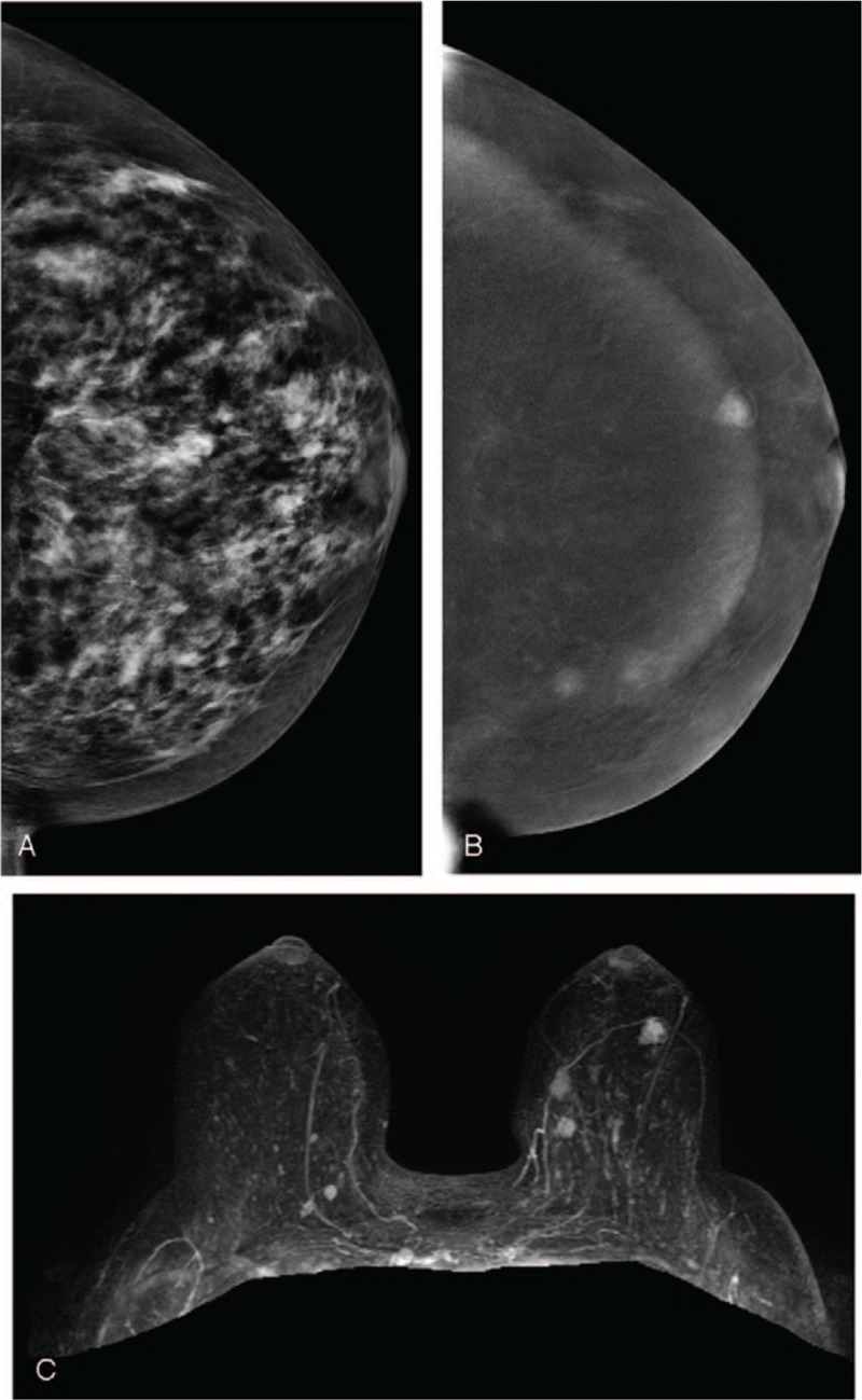
Figure 3.

A 50-year-old woman. (A) LE-MG (CC view) showed dense breasts without obvious breast nodules. (B) RSM (CC view) revealed three irregular enhanced masses in the inner region and subareolar region of the left breast. (C) CE-MRI with three-dimensional reconstruction displayed three enhanced masses corresponding to RSM. Finally, pathology confirmed that the two masses at the inner region were cancers and the mass at the subareolar region was a benign papillary tumor. CE-MRI = contrast-enhanced magnetic resonance imaging, LE-MG = low-energy mammography, RSM = recombine subtracted mammography.
