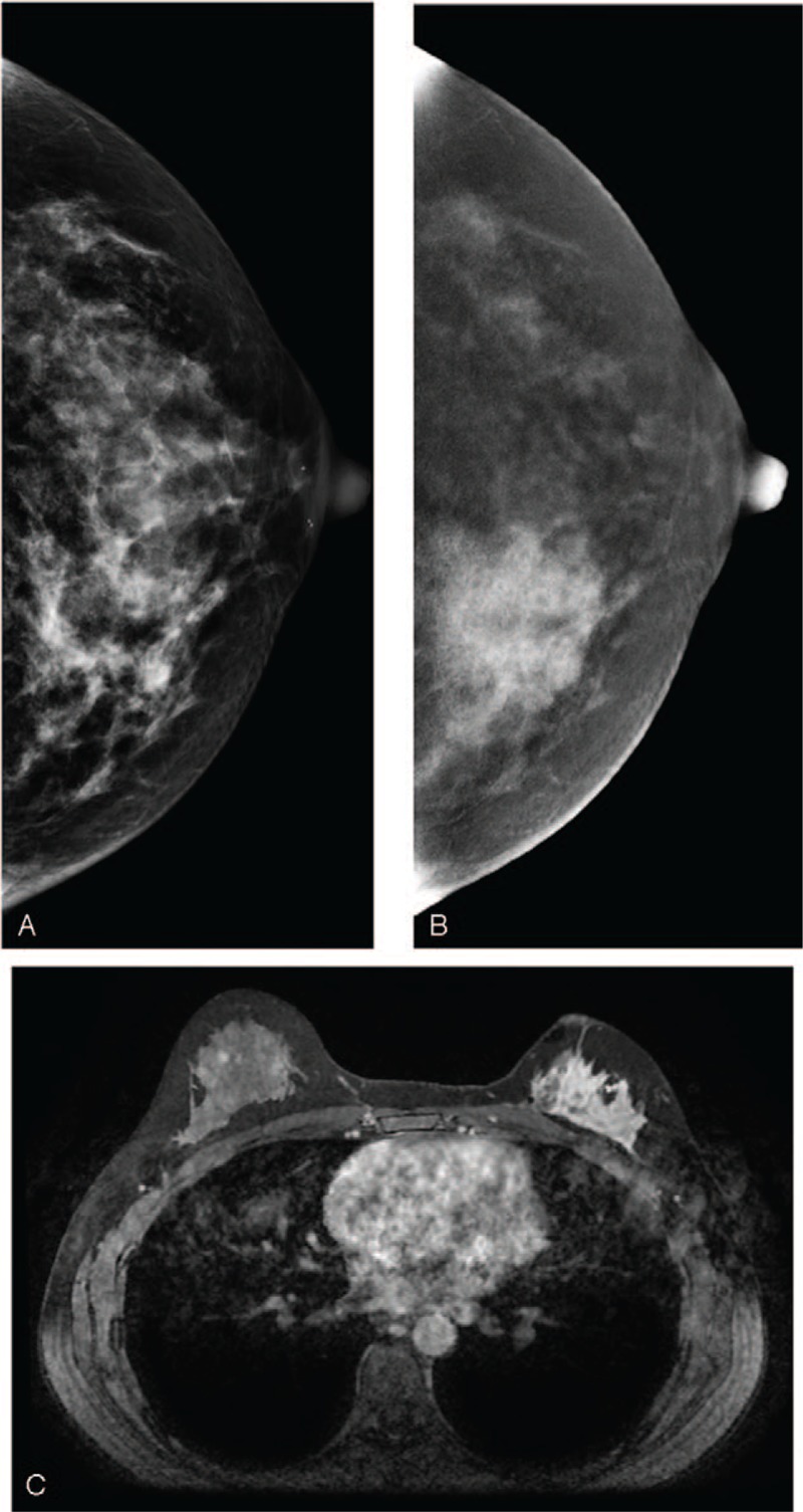
Figure 4.

A 38-year-old woman. (A) LE-MG (CC view) showed dense breasts with an irregular hyperdense patch of tissue distortion in the inner region of the left breast. (B) RSM (CC view) revealed a remarkable segmental enhancement in the inner region and multiple nodular enhancement in the outer region of the left breast. The patient requested partial mastectomy due to impalpable, negative sonography, and conventional mammography. The cancer was confirmed to be invasive ductal carcinoma. (C) CE-MRI 3 years after treatment demonstrated a non-mass enhanced recurrent cancer in the outer region of the left breast. CE-MRI = contrast-enhanced magnetic resonance imaging, LE-MG = low-energy mammography, RSM = recombine subtracted mammography.
